We're a community dedicated to beating blood cancer
Get InvolvedBlood Cancer UK
We research, we support, we care. Because it’s time to beat leukaemia, lymphoma, myeloma and all types of blood cancer.
How can we help?


Latest news and stories
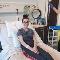
Omaze x Blood Cancer UK Research Fund launched
The research will look at new treatment options for people with myeloma, leukaemia and lymphoma
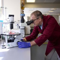
CAR-T therapy, Kymriah, available on NHS
The drug will be an option for young people with B-cell acute lymphoblastic leukaemia
Yescarta not recommended for use in Scotland
The blood cancer CAR-T therapy, Yescarta, is not to be made available on the NHS in Scotland for second line-use
Keep in touch with us
Get the latest news from Blood Cancer UK directly to your inbox.
We will keep you updated about our work and the ways you can help, including campaigns and events. We promise to respect your privacy and we will never sell or swap your details.